About the Melanoma
Melanoma is a serious form of skin cancer that begins in cells known as
melanocytes.
Melanoma skin cancer is more dangerous because of its ability to spread to other organs more rapidly
if it is not
treated at an early stage. Based on the global cancer data statistics, melanoma is among the twenty
most common cancer types in the world and when different types of skin cancers are considered,
melanoma can be identified as the deadliest form of skin cancer.
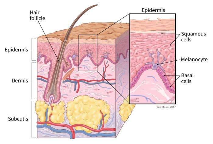
Melanocytes are skin cells found in the upper layer of skin. They produce a pigment known as
melanin, which gives skin its color.
When skin is exposed to ultraviolet (UV) radiation from the sun or tanning beds, it causes skin
damage that triggers the melanocytes to
produce more melanin, but only the eumelanin pigment attempts to protect the skin by causing the
skin to darken or tan.
Melanoma occurs when DNA damages from burning or tanning due to UV radiation which triggers
changes (mutations) in the melanocytes, resulting in uncontrolled cellular growth .
These mutations that occur in human DNA from UV radiation are called somatic mutations. On the other
hand, germline
mutations, or mutations that happen when cells are divided when producing offspring passes
hereditary diseases from parents to the child. It has been found that Melanoma can occur due to
both somatic and germline mutations in DNA.

Melanoma is present in many shapes, sizes and colors. That is why it is tricky to
provide a comprehensive set of warning signs. But it is vital to detect Melanoma at its early
stages and
start medical treatment so that it will not spread at a high rate and affect the other organs as
well which makes it difficult to be treated at its last stages. Melanoma is usually curable when
detected and treated early. Once melanoma has spread deeper into the skin or other parts of the
body, it becomes more difficult to treat and can be deadly.
At the beginning of the year 2021 it was
estimated that 207,390 cases of Melanoma will be diagnosed just in the U.S. and from
those patients
it was estimated that 7,180 people will die of Melanoma.
At present techniques like total cutaneous photography, dermoscopy, confocal scanning laser
microscopy (CSLM), ultrasound, magnetic
resonance imaging (MRI), optical coherence tomography (OCT), and multispectral imaging are being
used in
screening Melanoma of which dermoscopies are more widely being used.
When looking at the
statistics, it can be observed that fair-skinned people have a higher risk of being affected by
melanoma
when compared to darker-skinned people. The reason for this is that darker-skinned people
have more
eumelanin and fair-skinned people have more pheomelanin. As eumelanin can protect the skin from
being damaged by UV radiation from sources such as sun, tanning beds, etc. by darkening the
skin, fair-skinned people are more likely to be affected by melanoma due to lack of eumelanin.
Therefore, melanoma can be found commonly in European, American and Australian regions.
Melanoma Detection Tool

There were multiple reasons that made us create this online tool which could be used as an alternative to detect Melanoma in its earliest stages at a lesser cost and with a better accuracy so that the risk of the disease affecting the other organs is minimized . Studies have shown that a dermoscopy may even lower the diagnostic accuracy when being performed by inexperienced dermatologists, and therefore it needs to be performed by a dermatologist with a load of experience on skin lesions and an adequate amount of clinical practice. Even when an expert dermatologist uses a dermoscopy for diagnosis the accuracy level of it is estimated to be between 75-84%.
Even when the most common way of screening Melanoma is by using a dermoscopy , still in this
tool we
have included the option to use their DNA sequences as well to perform the diagnosis as there were
incidents where even though the person is affected with Melanoma, still it might take a while to be
noticeable on the surface of the skin.
As the first change occurs in the DNA, using the DNA sequence there are instances where Melanoma can
be detected at its very initial stages which makes the
medical procedures lesser and cure the patient sooner. Even a patient with a risk of being affected
by Melanoma due to a germline mutation, can use this tool to diagnose Melanoma from time to time so
that it could be detected as early as possible by detecting the mutations in the DNA sequence.
How the Tool Works
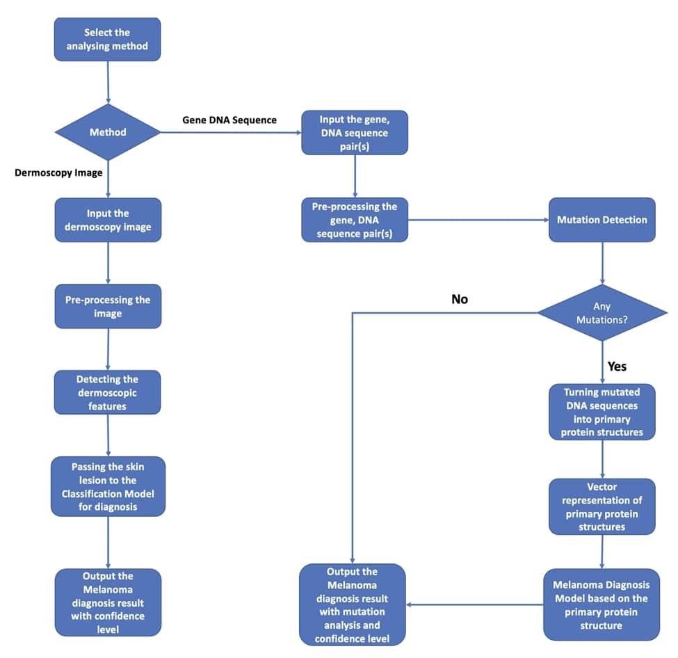
To start with, first you need to enter the general details requested from you to the portal and then select via which option you would prefer to perform a diagnosis on Melanoma using this online tool.
There are two options available in this tool namely by providing an image of the skin lesion or by providing the DNA sequence, from which you could select the most appropriate option based on the requirement.
If an image of the skin lesion is provided to the tool in order to perform the diagnosis, the tool will first execute the required pre-processing steps to obtain the image with better detail and less noise while segmenting the skin lesion from the background. Then the tool will look for the dermoscopic features that could be extracted from the image and based on those extracted dermoscopic features a trained classification model will output the diagnosis result with the level of confidence at which the diagnosis result was obtained.
If the other option is selected to perform the diagnosis based on the DNA sequence, the tool will first pre-process the DNA sequence and obtain the required genes from the sequence which causes Melanoma if mutated. Then the tool will find for any mutations in those genes and if no mutations were found, the tool will provide the output as Melanoma not detected.
But if any mutations are detected in this process, then the tool will convert the mutated DNA sequences to its primary protein structure and create a vector representation of the primary protein structure. Then using a trained model the diagnosis of Melanoma will be performed using the vector representation of the primary protein structure and the diagnosis result will be given as the output with the confidence level at which the diagnosis result was obtained.

Melanoma Diagnosis
Get in Touch


